Not on air · 2 of 3 networks checked

Stellaromics Inc. ads
- stellaromics.com
- Biotechnology Research
- US
- 65 employees
Stellaromics Inc.’s ads on LinkedIn, Meta and Google, recorded by adsrunning. Browse the latest here for free. Sign up free to see all 22. Watch Stellaromics Inc. on any plan to get each new one in your feed.
Watchlist included in every plan
What Stellaromics Inc. runs
- Single image · LinkedIn
- 2
- Video · LinkedIn
- 4
- Google ads on 10 Oct 2026
- 2
| Network | Status | Live ads | Nov 2025LinkedIn activity · 52 weeksNov 2026 | Checked | Next step |
|---|---|---|---|---|---|
| None recorded | 0 | Checked 3 Nov 2026 | See all 22 ads → | ||
| Meta | Not checked | — | |||
| Recently | 2 | Ads on the Google Ads Transparency Center | Checked 10 Oct 2026 | Collect them with the API → |
Stellaromics Inc. has no live LinkedIn ads. We recorded 2 Google ads on 10 Oct 2026.
Latest ads
- LINKEDIN · VIDEOSince 26 Oct 2026
🧬 What if the “neighbour” in your spatial map… isn’t actually a neighbour? 👀 In 2D, cells can overlap in XY….this creates “misinterpretation” or “misrepresentation” And when transcript signals sit near those overlapping boundaries, their cellular context can become ambiguous. 🧩 You can see the transcript. 🔬 But can you fully resolve its cellular context from XY alone? Then you add Z. 📐 Now you’re no longer looking at a dot on a plane. You can ask where that transcript sits within the tissue volume, which cellular volume surrounds it, and what cells are genuinely nearby. 🧠✨ A new 2026 preprint from Goode, Saunders and colleagues is a great example of where this is heading. 📄 Using STARmap-based in situ sequencing on the Pyxa platform in 50 µm intact mouse brain sections, the team acquired z-stack data and reconstructed RNA barcode decodes in three dimensions. 🧬🧠 The resulting data contained x, y and z positional information for individual decodes. 📍 So the question changes from: “Where is this transcript on the section?” to: “Where is this transcript in 3D, which cell surrounds it, and what is actually around that cell?” 🔄 3D isn’t just adding another axis. It’s adding biological context. 🌐 and that’s exactly what the Stellaromics Inc. Pyxa platform is built from the ground up for Because biology doesn’t happen in a plane. Your tissue isn’t 2D. Why should your spatial interpretation stop at XY? 🚀🔬 📄 Goode et al. (2026) Design and Assembly of Combinatorial DNA Barcodes for Probe-based Genomics Applications #SpatialBiology #SpatialTranscriptomics #3DSpatialBiology #SingleCell #Genomics
- LINKEDIN · IMAGESince 21 Oct 2026
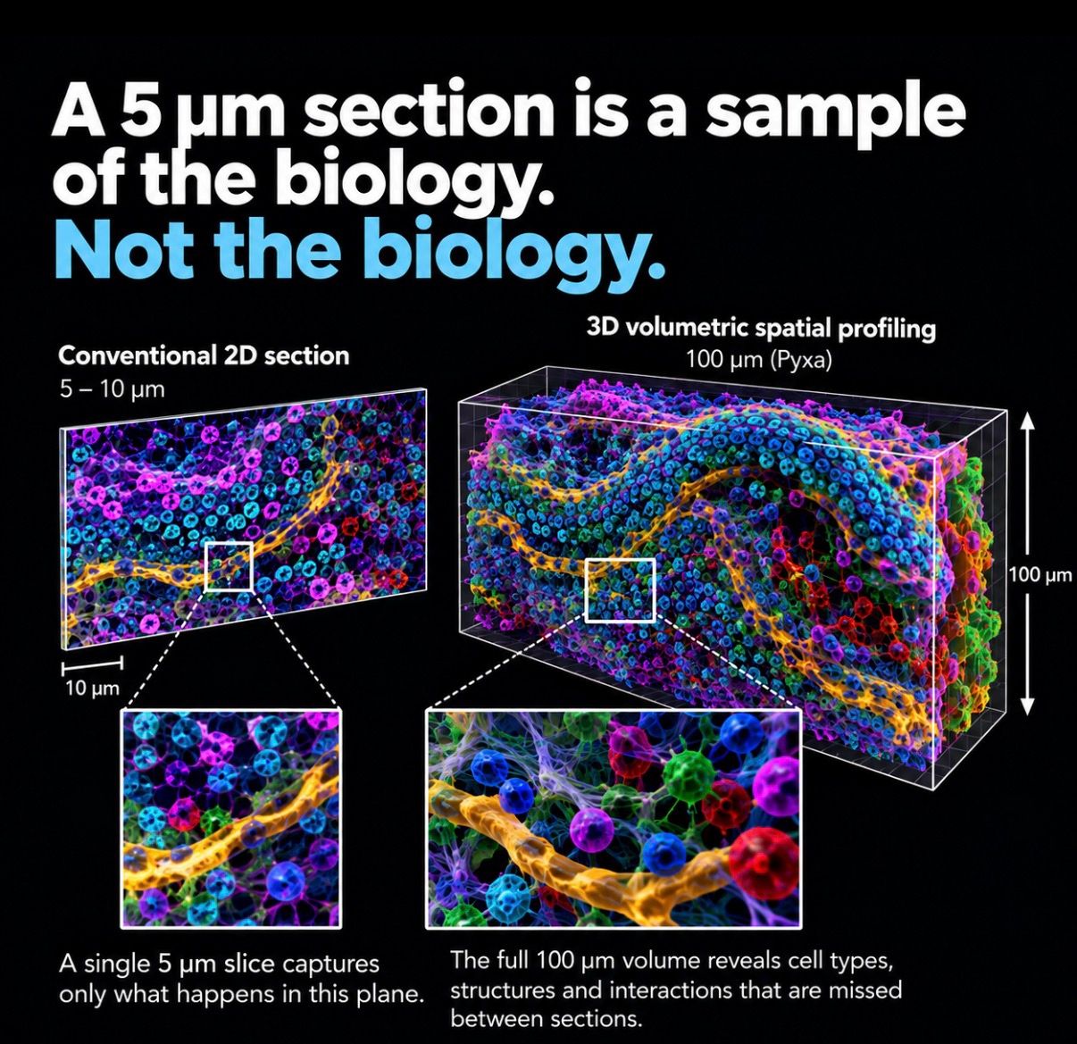
How much biology disappears when we cut the section? Cells don’t stop at the edge of a 5–10 µm slice. Neither do blood vessels, neuronal networks, tumour boundaries or cellular neighbourhoods. And neither do the interactions between them. This is why I find the move from spatial profiling in a plane to spatial profiling through tissue volume so interesting. A recent independent study provides a great example. Researchers used the Pyxa platform with STARmap chemistry in 50 µm mouse brain sections, acquiring z-stacks and reconstructing the three-dimensional distribution of RNA barcode decodes. It’s not simply about making a 3D image. It’s about preserving biological relationships that may never be visible in a single section. Same tissue. Same biology. A very different story when you can see what happens between the sections. 🔬 come and check it out with Stellaromics Inc. #SpatialBiology #SpatialTranscriptomics #3DSpatialBiology #SingleCell #Genomics #STARmap
- LINKEDIN · VIDEOSince 20 Oct 2026
Biological function depends on the interaction between cells and tissue structures in their native microenvironment: • tumor and immune cell interactions driving therapeutic resistance in cancer • microglia and amyloid-beta plaques promoting Alzheimer's disease progression • innate immune cells promoting epithelial barrier damage in inflammatory bowel disease In thin tissue slices, you can easily miss cell-cell interactions happening mere microns away from where you sectioned. 100 µm thick tissue sections capture 16x more cells, increasing the likelihood of capturing these events. What's hiding just beyond your slide? https://lnkd.in/e-PcGbz6/?utm_campaign=BoostedPosts-WebsiteVisits&utm_source=linkedin&utm_medium=paid&utm_id=1235771664&hsa_acc=545060280&hsa_cam=1235771664&hsa_grp=905105144&hsa_ad=1605792694&hsa_net=linkedin&hsa_ver=3
- LINKEDIN · IMAGESince 24 Sept 2026
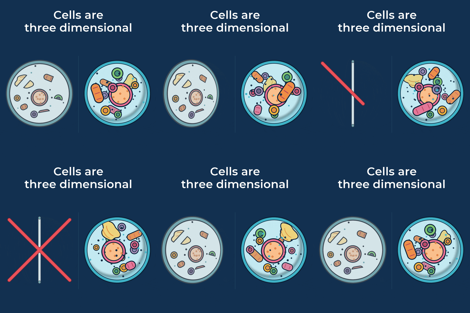
Cells are three-dimensional -- so why do we study them in flat tissue sections? Sectioning tissue down to 10 µm slices through cells, destroys cell morphology, and obfuscates the true 3D relationships between cells. In thick 100 µm tissue sections, intact layers of cells are preserved - maintaining complete cell and tissue architecture as well as the biologically-relevant relationships between cells in their native microenvironment. This has major implications for how we understand complex disease states such as neurodegeneration and cancer. Learn more about the advantages of 3D spatial multi-omics: https://lnkd.in/e5ksFyx7/?utm_campaign=Boost_Post_WebsiteVisits_Sep24,2026&utm_source=linkedin&utm_medium=paid&utm_id=1214336974&hsa_acc=538110497&hsa_cam=1214336974&hsa_grp=900386054&hsa_ad=1573884184&hsa_net=linkedin&hsa_ver=3
- LINKEDIN · VIDEOSince 23 Sept 2026
Thin tissue sections are prone to detecting overlapping, adjacent cells as a single cell during analysis. A recent publication showed that only 20-80% of cells in a 10 µm slice were consistently assigned the same cell type when the top and bottom 5 µm were analyzed separately - and the assignment rate varied greatly by region of the brain being analyzed. Does this worry you? Learn more about pivoting to true 3D spatial multi-omics, which allows you to capture multiple layers of intact cells in tissue sections up to 100 µm thick: https://lnkd.in/ewRXJc3x/?utm_campaign=Boost_Post_VideoViews_Sep23,2026&utm_source=linkedin&utm_medium=paid&utm_id=1214263424&hsa_acc=538110497&hsa_cam=1214263424&hsa_grp=900198324&hsa_ad=1573439534&hsa_net=linkedin&hsa_ver=3
- LINKEDIN · VIDEOSince 21 Aug 2026
Most spatial platforms can only image 5–10 µm of tissue. Pyxa® goes to 100 µm. Stellaromics built Pyxa® for True 3D™ spatial multi-omics: volumetric imaging of intact tissue up to 100 µm thick, subcellular localization of hundreds of mRNA transcripts, and xyz coordinates for every cell and transcript in the sample. Pyxa is built on STARmap and provides an end-to-end workflow from kit-based sample prep through automated fluidics and confocal imaging to analysis in PyxaStudio™. The result: cell-cell relationships measured in the volume they actually occupy, rather than projected onto a plane. Why 3D? Download the white paper to learn more.
6 of 22 adsSign up to see every Stellaromics Inc. ad, filter by format and date, and get alerts on new ones.
See all 22 adsQuestions about Stellaromics Inc.’s ads
Is Stellaromics Inc. running LinkedIn ads?
We recorded no live LinkedIn ads for Stellaromics Inc. on 7 Nov 2026.
How many LinkedIn ads has Stellaromics Inc. run?
LinkedIn’s ad library lists 22 ads for Stellaromics Inc.. We recorded 22 LinkedIn ads for Stellaromics Inc., first recorded on 1 Oct 2026 and last recorded on 28 Oct 2026.
Does Stellaromics Inc. run Google ads?
Yes. We recorded 2 Google ads for the domain stellaromics.com on 10 Oct 2026.
Which ad formats does Stellaromics Inc. use?
The 6 LinkedIn ads on this page include 2 image ads and 4 video ads.
Watch the companies you care about.
Filter every ad by industry, country, format and network, and get alerts when a company you watch starts running new ads.