On air · 1 of 3 networks
AIRS Medical
- airsmed.com
- Healthcare & Life Sciences
- KR
- 162 employees
AIRS Medical’s ads on LinkedIn, Meta and Google, recorded by adsrunning. Browse the latest here for free. Sign up free to see all 517. Watch AIRS Medical with Starter to get each new one in your feed.
Watchlist with Starter · $49/mo
| Network | Status | Live ads | Nov 2025LinkedIn activity · 52 weeksOct 2026 | Checked | Next step |
|---|---|---|---|---|---|
| Running | 81 | Checked 30 Oct 2026 | See all 517 ads → | ||
| Meta | Not checked | — | |||
| Not checked | — |
AIRS Medical has 81 live LinkedIn ads.
Latest ads
- LINKEDIN · IMAGESince 1 Sept 2026
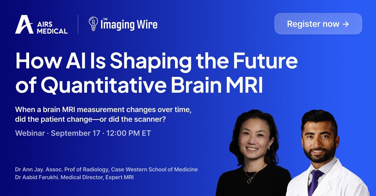
When a brain MRI measurement changes over time, did the patient change—or did the scanner? Join Dr. Ann Jay, MD, FACR, Assistant Professor of Radiology, Case Western School of Medicine Associate Professor of Radiology, Case Western School of Medicine and Dr. Aabid Farukhi, MD, Medical Director, Expert MRI clinical discussion on how AI is helping address this challenge. What you’ll learn: ✅ How to improve consistency in quantitative measurements across scanners, protocols, and field strengths ✅ How AI-powered segmentation can go beyond standard volumetry ✅ How longitudinal analysis can help identify meaningful patient change over time ✅ How these capabilities are being applied to real patient cases and clinical workflows Who should attend? Neuroradiologists • Radiologists • Imaging Directors • MRI Program Leaders • Clinical Imaging Teams • AI & Imaging Innovation Leaders Why attend? If distinguishing true patient change from imaging variability is important to your work, you won’t want to miss this discussion. 👉 Register now: https://lnkd.in/eN7QQ3PZ/?utm_campaign=InsightWebinarWebsiteConversions&utm_source=linkedin&utm_medium=paid&utm_id=1206685974&hsa_acc=517791495&hsa_cam=1206685974&hsa_grp=887234784&hsa_ad=1541221004&hsa_net=linkedin&hsa_ver=3 Want to learn more before the webinar? Reach out to Jina Park, Chief Strategy Officer at AIRS Medical, to learn more about SwiftSight and how it can support consistent quantitative brain MRI across your imaging environment. #SwiftSight #BrainMRI #Neuroimaging #QuantitativeMRI #Radiology #AIinRadiology #AIRSMedical
- LINKEDIN · VIDEOSince 10 Jul 2026
For Chiron Group, faster MRI is not just about speed. Dr. Andrea Cicchiello, President and Founder explains how SwiftMR® helps imaging providers get more from their existing MRI systems—saving time, improving patient flow, and strengthening day-to-day financial performance. Watch the video to hear Chiron Group’s perspective on the operational value of faster MRI.
- LINKEDIN · VIDEOSince 10 Jul 2026
What did a 36% reduction in MRI scan time mean for Sanitas? After implementing SwiftMR®, Sanitas increased patient volume by eight patients per day while also improving image quality—without replacing its existing MRI equipment. The result: greater daily capacity, more efficient MRI operations, and shorter examinations for patients. Watch the video to hear how Sanitas is getting more from its existing MRI system with SwiftMR.
- LINKEDIN · IMAGESince 15 Apr 2026
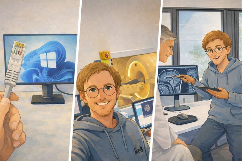
𝗪𝗵𝗮𝘁 𝗿𝗲𝗮𝗹𝗹𝘆 𝗵𝗮𝗽𝗽𝗲𝗻𝘀 when you upgrade your MRI with AI A look behind the scenes with 𝗦𝘄𝗶𝗳𝘁𝗠𝗥 What does implementation actually look like, and 𝗵𝗼𝘄 𝗹𝗼𝗻𝗴 𝘄𝗶𝗹𝗹 𝗺𝘆 𝗠𝗥𝗜 𝗯𝗲 𝗱𝗼𝘄𝗻? Here is a real-world example. 𝗗𝗮𝘆 𝟭 15:00 – Arrival of the SwiftMR Customer Success Manager 15:15 – Coffee (important step) 15:30 – Integration of the SwiftMR gateway / virtual machine with the 𝗠𝗥𝗜 𝗮𝗻𝗱 𝗣𝗔𝗖𝗦 17:00 – Optimization of MRI sequences (2–3 hours) 𝗗𝗮𝘆 𝟮 08:00 – Normal patient scanning with 𝗔𝗜-𝗮𝗰𝗰𝗲𝗹𝗲𝗿𝗮𝘁𝗲𝗱 𝘀𝗲𝗾𝘂𝗲𝗻𝗰𝗲𝘀 11:00 – Image review and discussion with the radiologist ✔ 𝗜𝗻𝘀𝘁𝗮𝗹𝗹𝗮𝘁𝗶𝗼𝗻 𝗰𝗼𝗺𝗽𝗹𝗲𝘁𝗲𝗱 𝗧𝗵𝗲 𝗸𝗲𝘆 𝘁𝗮𝗸𝗲𝗮𝘄𝗮𝘆: A simple and straightforward installation with only 𝗼𝗻𝗹𝘆 𝟮–𝟯 𝗵𝗼𝘂𝗿𝘀 𝗼𝗳 𝗠𝗥𝗜 𝗱𝗼𝘄𝗻𝘁𝗶𝗺𝗲. How do you obtain the best result from sequence optimization? When integrating AI into MRI, 𝗿𝗮𝗱𝗶𝗼𝗹𝗼𝗴𝗶𝘀𝘁 𝗳𝗲𝗲𝗱𝗯𝗮𝗰𝗸 𝗶𝘀 𝗰𝗿𝗶𝘁𝗶𝗰𝗮𝗹. Image quality is not a fixed target—every radiologist has different expectations and clinical priorities. That is why iterative improvement and fine-tuning matter. One way to think about AI in MRI is this: AI gives your MRI additional signal. The decision where to use it is 𝗳𝘂𝗹𝗹𝘆 𝗶𝗻 𝘆𝗼𝘂𝗿 𝗵𝗮𝗻𝗱𝘀. Do you want: – Higher resolution – Faster scans – Thinner slices Or a balance of all three? You decide where to use the additional signal—and 𝗴𝗲𝘁 𝘁𝗵𝗲 𝗺𝗼𝘀𝘁 𝗼𝘂𝘁 𝗼𝗳 𝘆𝗼𝘂𝗿 𝘀𝘆𝘀𝘁𝗲𝗺.
- LINKEDIN · IMAGESince 15 Apr 2026
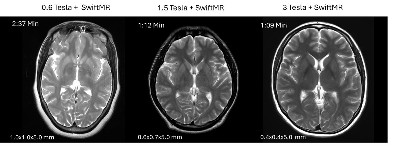
𝗜𝘀 𝗮 𝟯 𝗧𝗲𝘀𝗹𝗮 𝗠𝗥𝗜 𝗿𝗲𝗮𝗹𝗹𝘆 𝗯𝗲𝘁𝘁𝗲𝗿 𝘁𝗵𝗮𝗻 𝗮 𝟭.𝟱 𝗧𝗲𝘀𝗹𝗮 𝗠𝗥𝗜? You’ve probably asked yourself that question. Yes—3T offers higher signal. But it also introduces real trade-offs: 𝗤𝘂𝗶𝗰𝗸 𝗮𝗻𝗮𝗹𝘆𝘀𝗶𝘀: 𝗦𝗡𝗥: Theoretically, SNR can double, but real-world gains are often closer to ~30–60% depending on anatomy, coils, and protocol. This can be translated into higher resolution and/or faster scans. 𝗦𝗔𝗥: SAR can increase by up to ~4× at 3T (sequence- and patient-dependent). You often need to compensate with other parameters, e.g., reduced flip angle, longer TR, or different refocusing strategies. 𝗦𝘂𝘀𝗰𝗲𝗽𝘁𝗶𝗯𝗶𝗹𝗶𝘁𝘆 / 𝗱𝗶𝗲𝗹𝗲𝗰𝘁𝗿𝗶𝗰 𝗲𝗳𝗳𝗲𝗰𝘁𝘀: These effects are generally more pronounced at 3T and are often negative in practice (e.g., more artifacts, greater sensitivity to implants, B1 inhomogeneity—especially in body imaging). 𝗦𝗽𝗲𝗰𝘁𝗿𝗼𝘀𝗰𝗼𝗽𝘆: 3T clearly wins here due to better peak separation at higher field strength. 𝗪𝗵𝗮𝘁 𝘁𝗵𝗮𝘁 𝗺𝗲𝗮𝗻𝘀 𝗳𝗼𝗿 𝘆𝗼𝘂 𝗮𝘀 𝗮 𝗰𝗹𝗶𝗻𝗶𝗰𝗶𝗮𝗻: 3T imaging comes with meaningful downsides—particularly for abdominal imaging—where SAR limits, B1 inhomogeneity, and artifacts can reduce robustness. In many cases, you may be better off investing in: State-of-the-art coils that improve SNR and enable more parallel imaging acceleration. AI-based image enhancement (e.g., SwiftMR), which can boost effective SNR by roughly ~1.4×, bringing you closer to 3T-like performance for many applications. This can give you a solid workhorse system that delivers high-quality images with fast scan times. If you need truly small voxels (e.g., in neuro/brain imaging or other specialized applications), the additional SNR from a 3T system can be beneficial—especially when combined with AI. 𝗠𝘆 𝘁𝗮𝗸𝗲: Keep what works, upgrade intelligently, and be strategic. For years, the field has pushed toward higher field strengths. More recently, vendors have introduced new MRI models below 1.5T. This isn’t about 3T replacing 1.5T or low-field “taking over.” It’s about understanding the pros and cons of each field strength and choosing what fits your use case. No matter the field strength, AI-based image enhancement is increasingly key to keeping your system state-of-the-art. images shown below illustrate the different field strengths. 3T could be even faster at same resolution.
- LINKEDIN · IMAGESince 15 Apr 2026
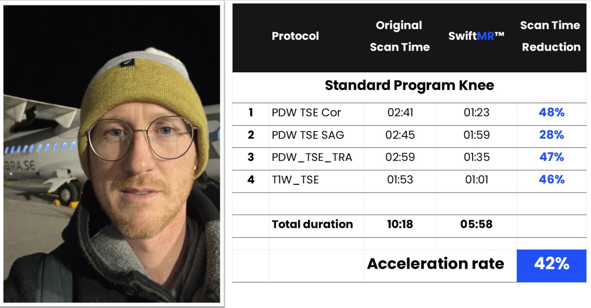
𝗙𝗮𝘀𝘁𝗲𝗿 𝗠𝗥𝗜 𝗮𝗻𝗱 𝗕𝗲𝘁𝘁𝗲𝗿 𝗜𝗺𝗮𝗴𝗲 𝗤𝘂𝗮𝗹𝗶𝘁𝘆 — 𝗖𝗮𝗻 𝗬𝗼𝘂 𝗛𝗮𝘃𝗲 𝗕𝗼𝘁𝗵? I just returned to Germany for another SwiftMR installation at a customer site. Almost every conversation starts with the same request: “Can we scan faster and still improve image quality?” Traditionally, MRI acceleration comes at a cost—either reduced SNR or compromised spatial resolution. That trade-off has long been accepted as unavoidable. However, this changes when AI-based image enhancement is used to recover SNR. With additional SNR made available by AI, you can decide how to spend it: • 𝗣𝘂𝗿𝗲 𝗮𝗰𝗰𝗲𝗹𝗲𝗿𝗮𝘁𝗶𝗼𝗻 – achieve up to 50% scan-time reduction without sacrificing image quality • 𝗕𝗮𝗹𝗮𝗻𝗰𝗲𝗱 𝗼𝗽𝘁𝗶𝗺𝗶𝘇𝗮𝘁𝗶𝗼𝗻 – combine acceleration with higher spatial resolution, choosing the right mix for your clinical need • 𝗛𝗶𝗴𝗵𝗲𝗿 𝗿𝗲𝗰𝗲𝗶𝘃𝗲𝗿 𝗯𝗮𝗻𝗱𝘄𝗶𝗱𝘁𝗵 – reduce distortion and susceptibility artifacts, particularly in challenging anatomy 𝗠𝘆 𝘁𝗮𝗸𝗲𝗮𝘄𝗮𝘆: Its up to you whether to go for speed or use the additional SNR from AI for resolution increase. If you increase resolution and scan time to much, longer high-resolution scans actually perform worse than faster, accelerated acquisitions. Patient motion increases with scan time, leading to blurring that potentially negates any theoretical resolution gain. So the shorter scan may deliver the sharper image. My recommendation: run side-by-side comparisons on a few patients. Verify whether your longer protocol truly improves resolution—or whether the fast scan produces superior diagnostic quality especially in difficult patients. 𝗧𝗵𝗲 𝗿𝗲𝘀𝘂𝗹𝘁 𝗳𝗿𝗼𝗺 𝘁𝗵𝗶𝘀 𝘃𝗶𝘀𝗶𝘁: • Knee protocol accelerated by 42% • Lumbar spine scan time reduced by 49% What was the biggest challenge of this installation? Clearly that the airline left my luggage behind in Copenhagen 😅
6 of 517 adsSign up to see every AIRS Medical ad, filter by format and date, and get alerts on new ones.
See all 517 adsWatch the companies you care about.
Filter every ad by industry, country, format and network, and get alerts when a company you watch starts running new ads.